
Correction of the Supratip Deformity of the Nose
| 2025-07-01
Learn causes, surgical techniques, and outcomes for correcting supratip deformity after rhinoplasty with effective approaches and high patient satisfaction.

Seok-Kwun Kim, MD, PhD, Ju-Chuan Kim, MD, Keun-Cheol Lee, MD, PhD, Hyun-Soo Kim, MD, PhD
Dr Seok-Kwun Kim, Dr Ju-Chan Kim, and Dr Lee are from the
Department of Plastic and Reconstructive Surgery, Dong-A
University Hospital, Busan, Korea. Dr Hyun-Su Kim is from Nose
Plastic Surgery Clinic, Busan, Korea.
Aesthetic Surgery Journal, Volume 32, Issue 8, November 2012, Pages 943–955, https://doi.org/10.1177/1090820X12463386
Corresponding Author:
Dr Seok-Kwun Kim, Department of Plastic and Reconstructive
Surgery, Dong-A University School of Medicine, 26, Daesingongwon-
Ro, Seo-Gu, Busan, 602-715, Korea.
E-mail: sgkim1@dau.ac.kr
Published: 01 November 2012
Abstract
Background: A supratip deformity can develop either congenitally or after a poorly executed rhinoplasty, as a result of fullness or convexity just above the nasal tip.
Objective: In this article, the authors describe the causes of the supratip deformity and present their technique for surgical correction.
Methods: A retrospective chart review was conducted for 62 consecutive patients (24 primary rhinoplasty and 38 secondary rhinoplasty) treated between January 2005 and February 2010. The authors’ approach to managing supratip deformity included a combination of wide undermining of the nasal skin through the open technique; resection of excessive soft tissue and the bony, cartilaginous dorsum; suturing to advance the supratip; and augmentation of the nasal tip and dorsum with cartilage, crushed cartilage, silicone implants, and/or septal extension grafts.
Results: No infection or nasal bleeding was recorded during postoperative follow-up. The supratip and tip projection showed adequate shape in all cases. To assess satisfaction, all patients and 2 independent plastic surgeons were asked to rate the result on a 5-point scale. The average patient satisfaction rating was 4.2 and the average independent rating was 4.4 points.
Conclusions: Supratip deformities should be corrected according to their unique cause and shape. The authors’ method of repairing these deformities has resulted in a low complication rate and yielded satisfactory results.
Level of Evidence: 4
Keywords
: supratip deformity, supratip advancement suture, septal extension graft, rhinoplasty
INTRODUCTION
Supratip deformity of the nose is one of the most common postoperative deformities that requires secondary rhinoplasty [1-4]. The supratip deformity, previously called the “pollybeak” or “parrot beak” deformity, is defined as fullness or convexity immediately cephalad to the nasal tip, which may occur as a primary (congenital) or a secondary (postrhinoplasty) deformity [5,6]. Primary supratip deformity is characterized by an overprojected supratip, an underprojected tip, and a cephalic-oriented lower lateral cartilage. A secondary supratip deformity is caused by supratip soft tissue excess, an overresected supratip, residual excess in the caudal dorsum, inadequate tip projection, and a residual, excessive septum of the supratip and an underprojected tip (Table 1). In this article, we describe our method of addressing a supratip deformity, which accounts for the causes and shape of each patient’s individual anatomy, and present the surgical results from a case series of patients treated with this approach.
| Table 1. Classification of Supratip Deformity |
|---|
| Primary supratip deformity |
| Overprojected supratip (caudal dorsum) |
| Underprojected tip |
| Cephalically oriented lower lateral cartilage |
| Secondary supratip deformity |
| Supratip soft tissue excess |
| - Early postopertive period (1-3 months) |
| - Late postoperative period (after 1 year) |
| Overresected supratip (soft tissue excess) and inadequate tip projection |
| Residual excessive caudal dorsum |
| Inadequate tip projection |
| Residual excessive caudal septum and underprojected tip |
METHODS
We conducted a retrospective review of 62 consecutive patients with supratip deformity (24 primary and 38 secondary) treated between January 2005 and February 2010.
Operative Technique
Each patient’s deformity was defined through a detailed history and complete aesthetic nasal analysis, and the etiology was identified.
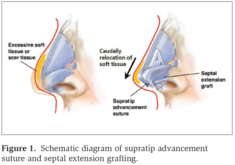
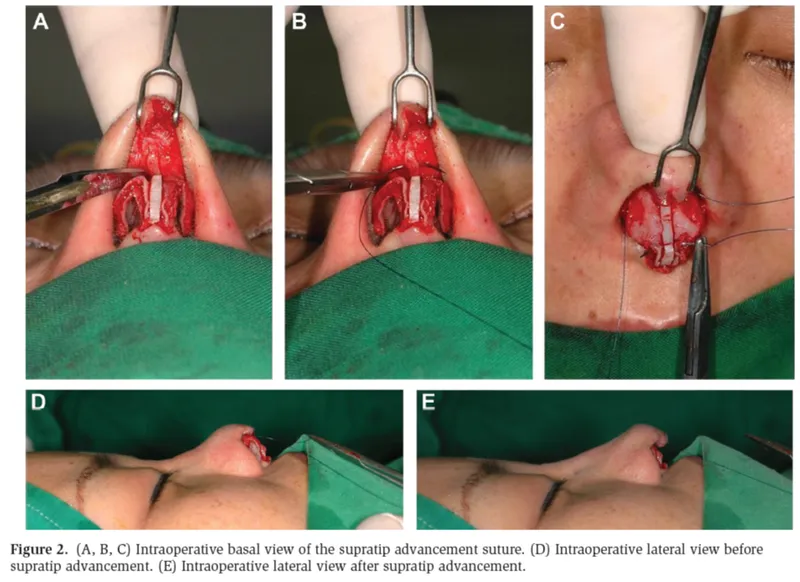
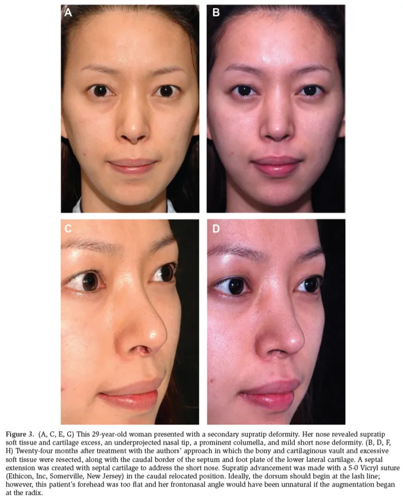
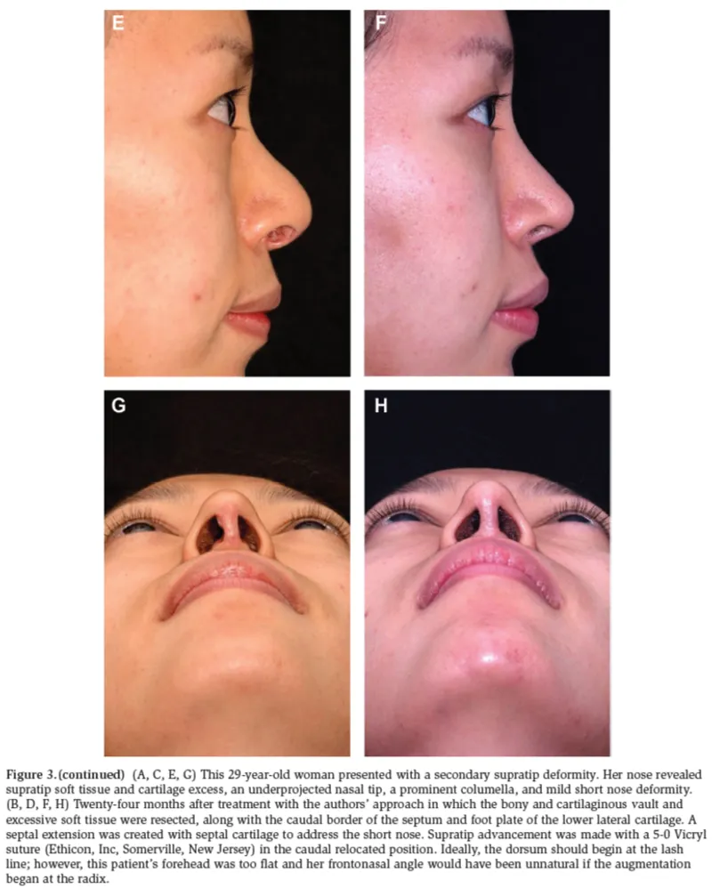
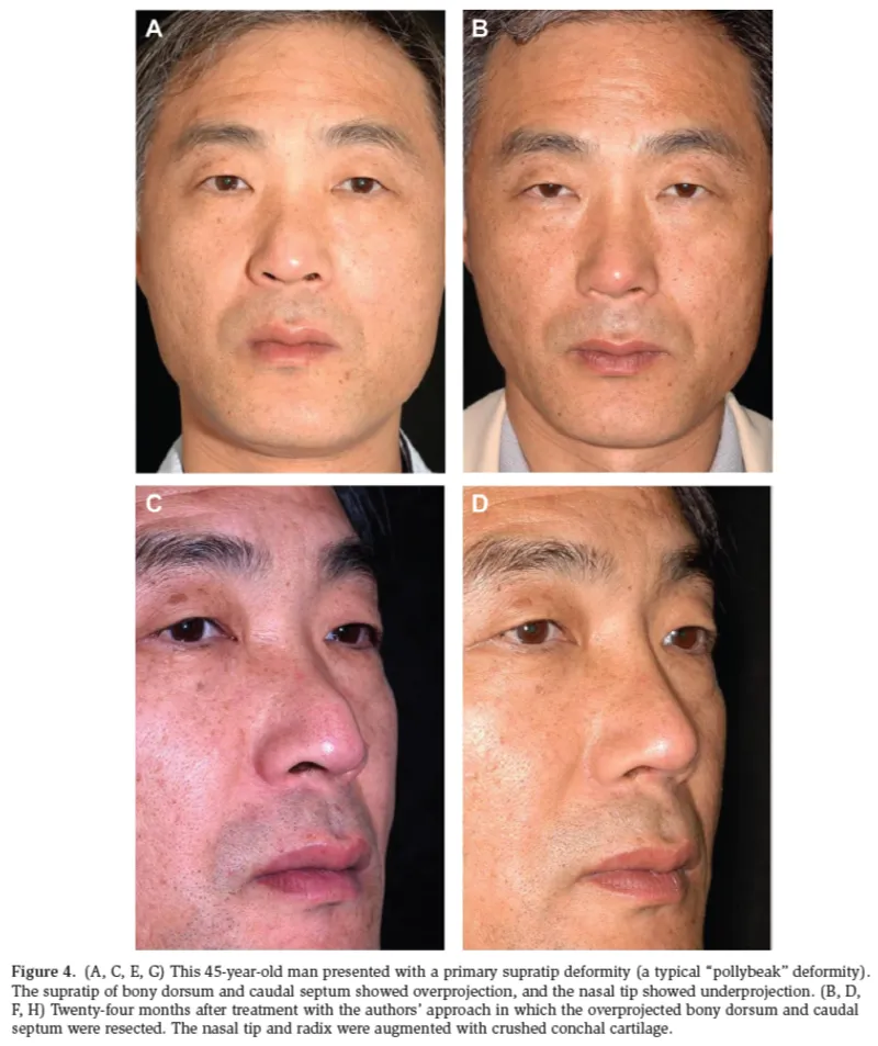
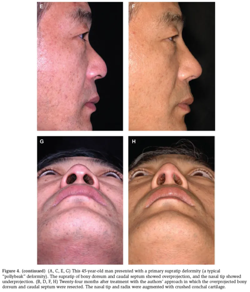
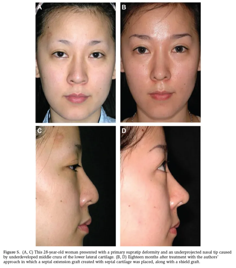
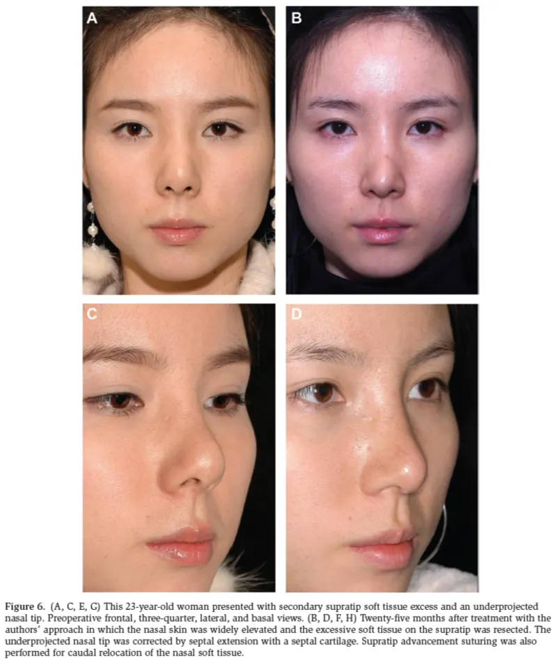
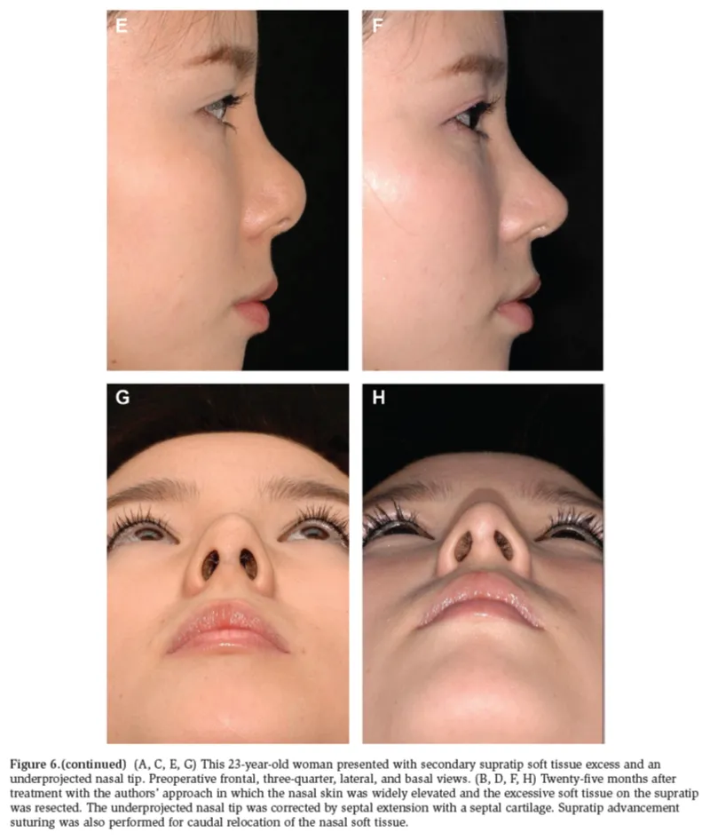
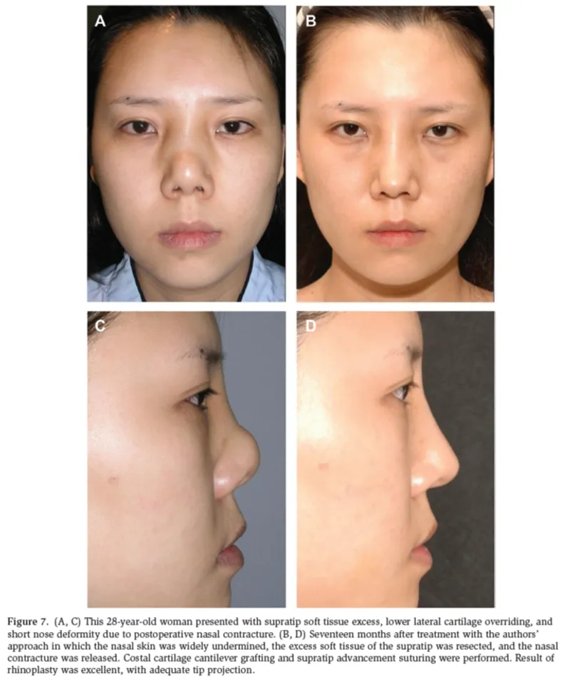
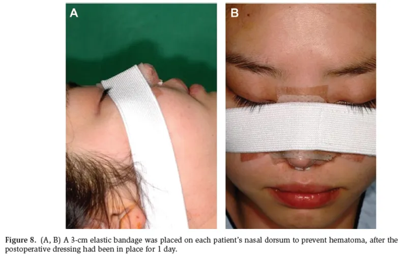
The open rhinoplasty technique was used; each patient received local anesthetic solution (1% lidocaine with 1:100,000 epinephrine). Although the method of correction did not differ significantly based on whether the patient had a primary or secondary deformity, certain aspects of the approach were tailored to each patient’s anatomy. Our principal management of the supratip deformity was a combination of (1) wide undermining of the nasal skin through open rhinoplasty, (2) resection of the excessive soft tissue and bony and cartilaginous dorsum, (3) use of a supratip advancement suture (Figures 1 and 2), (4) augmentation of the nasal tip and dorsum with cartilage or crushed cartilage, and (5) a septal extension graft using septal cartilage or rib cartilage (Figure 1). Specific operative details for selected patients are given in Figures 3 to 7. Postoperatively, we applied a 3-cm elastic bandage to the nasal dorsum to prevent hematoma, after dressing for 1 day (Figure 8).
RESULTS
There were 13 men (21%) and 49 women (79%) in our series of 62 patients; their average age was 35.6 years. The 24 cases of primary supratip deformity included 12 cases of overprojected supratip, 9 cases of underprojected tip, and 3 cases of cephalically oriented lower lateral cartilages. Of the 38 cases of secondary supratip deformity, there were 10 cases of recalcitrant soft tissue excess, 8 cases of overresected caudal dorsum, 9 cases of residual excessive caudal dorsum, 8 cases of underprojected tip, and 8 cases of residual excessive caudal septum. Most patients with a supratip deformity had more than one causative factor.
No infection or nasal bleeding was recorded during postoperative follow-up. Antibiotics (Tiroxin, 250 mg, 3 times a day) were administrated orally in all cases for 5 days postoperatively. A nasal splint and butterfly stitches were placed intraoperatively and were removed 5 days postoperatively. The follow-up period ranged from 1 to 3 years. No other supratip deformity was noted at follow-up evaluation in any patient. The supratip and tip projection showed adequate shape in all patients.
Results were assessed by a subjective satisfaction rating from the patients and 2 independent plastic surgeons. For evaluation of subjective satisfaction, patients were directly asked to assess their satisfaction on a 5-point scale after the procedure: (1) no effective correction of supratip deformity (1 point), (2) disharmony of the supratip and tip of the nose (2 points), (3) acceptable nasal shape but some degree of remnant supratip deformity (3 points), (4) relatively good correction of the supratip and tip (4 points), and (5) very satisfactory correction of the supratip and nasal shape (5 points). The average patient satisfaction rating was 4.2.
For the independent evaluation portion, a series of preoperative and postoperative photographs were given to 2 plastic surgeons; these photos included a frontal view, three-quarter oblique view, lateral view, and basal view. The surgeons evaluated 5 parameters: (1) adequacy of tip projection, (2) adequacy of caudal dorsal projection, (3) presence of supratip break, (4) suitability of the amount of soft tissue in the supratip area, and (5) adequacy of the lower lateral cartilage change and septal extension. Each category was allowed up to 1 point; thus, the total potential score was 5 points. The average score was 4.4, which was regarded as a highly successful correction.
Clinical results are shown in Figures 4 to 7.
DISCUSSION
A supratip deformity reveals fullness or convexity just above the nasal tip. It is not the transient supratip swelling that occurs in the immediate postoperative period; it is a definite and persistent deformity [7]. Guyuron et al. [8] described the supratip deformity as a hallmark of a poorly executed rhinoplasty or inauspicious healing. Clinically, 9% of primary rhinoplasty patients demonstrated supratip fullness [8]. Many Asians have primary, as well as secondary, supratip deformities. In these cases and many others, management of primary and secondary supratip deformity does not differ drastically, although etiology is important in developing an operative plan: Are displaced anatomic structures present? Have structural components been underresected or overresected? Has a combination thereof occurred? By answering these questions, appropriate surgical goals and treatment plans can be established. First, displaced anatomic structures should be repositioned appropriately. Then, areas of underresection should be identified. Likewise, in areas of overresection, missing tissue should be identified and replaced as needed. Finally, the optimal method of approach is determined based on the overall findings and the deformity itself [9].
A pollybeak is one of the most common deformities after primary rhinoplasty; most studies suggest that the condition occurs in 40% to 64% of cases for which secondary rhinoplasty was performed. The appearance of a pollybeak is created when the supratip projects beyond the tip in the plane of the nasal dorsum. This gives the nose a bulbous, rounded shape with the illusion of tip ptosis due to the absence of a supratip break [4,10,11]. For patients who had an overprojected supratip or supratip soft tissue excess and excessive residual caudal dorsum, resection of the soft tissue, residual nasal bone, and upper lateral cartilage was needed, but no single technique has been adopted to correct supratip deformity. Jung et al. [6] performed an elliptical supratip skin excision to reduce the extra soft tissue and to lower the supratip area at the risk of an external scar. In their study, the small linear scar that occurred as a result of this procedure consistently faded after several months and was aesthetically unremarkable. Hanasono et al. [5] used triamcinolone injection for correction of the soft tissue pollybeak deformity. Triamcinolone was injected at 1 week after surgery, and a secondary injection was performed at 4 weeks after surgery. Eighty-five percent of patients had acceptable results, with good supratip definition. Campus et al. [12] used “the trapezoid peak” technique to prevent supratip deformity in aesthetic rhinoplasty. This technique is a simple and reliable septal refinement for patients requiring extratip support but not grafts and may be useful in preventing loss of tip projection and supratip deformity. Rees et al. [13] suggested trimming the high dorsum, excising scar tissue, and lowering the upper lateral cartilages, as necessary. Although they recognized the effectiveness of increasing tip projection through columellar advancement and alar cartilage modification, they advised against the use of alloplastic materials or autogenous grafts because of the risk for extrusion or resorption. As stated, our management of the supratip deformity includes a combination of wide undermining of the nasal skin through the open rhinoplastic technique; resection of the excessive soft tissue and the bony, cartilaginous dorsum; use of a supratip advancement suture; augmentation of the nasal tip and dorsum with cartilage or crushed cartilage; and a septal extension graft using a septal or rib cartilage.
With cases of underprojected tip or inadequate projection, tip projection must be increased by augmentation, septal extension cartilage grafts, spreader grafts, and mini spreader grafts [14,15]. For cases of underprojected tip, Guyuron et al. [8] recommended tip grafts for small lobule and columellar struts, for an adequate lobule. For an underprojected midvault, they proposed the use of septal, costochondral, or conchal cartilage grafts. For cephalically oriented lower lateral cartilages, they suggested simply repositioning them to address supratip prominence. Finally, they supported Sheen’s [7] concept that most supratip deformities are the result of overzealous resection and that the deformity is the product of scar tissue formed in response to overresection and creation of dead space.
In summary, for correction of an overresected supratip and inadequate tip projection, cartilage grafting, tip augmentation, and septal extension grafting are recommended. In addition, implant material such as expanded polytetrafluoroethylene or silicone can sometimes also be used in Asian individuals. For a patient with residual excessive septum of the supratip and an underprojected tip, tip augmentation and resection of the caudal dorsum can be recommended. Direct contact between the subcutaneous portion of the skin flap and the cartilage is very important in repairing a supratip deformity. Advancement suturing of the nasal soft tissue is also a very useful technique. Scar tissue that develops as a result of the supratip manipulation can reduce dead space, and the nasal tip can be relocated by use of this technique. Septal extension grafting using septal, conchal, or rib cartilage is recommended in a patient with a short nose combined with an underprojected tip [14].
Nasal skin necrosis has been reported after supratip advancement suturing; however, in the current series, there were no cases of skin necrosis because the series included patients with thick soft tissue at the supratip. Review of the comprehensive literature describing both the supratip deformity and its correction suggests an approach tailored not only to diagnosing the problem but also to identifying its etiology. In the end, the best method for preventing a secondary supratip deformity is to avoid its etiologic factors. This can be accomplished only through a systemic preoperative analysis of the nasal architecture and a focused surgical plan [16].
CONCLUSIONS
Supratip deformities should be corrected in accordance with their etiology and the shape of the deformity. Our management of the supratip deformity includes a combination of wide undermining of the nasal skin through the open rhinoplastic technique; resection of the excessive soft tissue and bony, cartilaginous dorsum; supratip advancement suturing; augmentation of the nasal tip and dorsum with cartilage or crushed cartilage; and septal extension grafting with septal, conchal, or rib cartilage. This approach showed satisfactory results with both primary and secondary supratip deformities of the nose.
Disclosures
The authors declared no potential conflicts of interest with respect to the research, authorship, and publication of this article.
Funding
This study was supported by a research grant from Dong-A University.
REFERENCES
- Parkes ML, Kanodia R, Machida BK. Revision rhinoplasty: an analysis of aesthetic deformities. Arch Otolaryngol Head Neck Surg. 1992;118:695-701.
- O’Connor GB, McGregor MW. Secondary rhinoplasties: their cause and prevention. Plast Reconstr Surg (1946). 1955;15:404-410.
- Swanepoel PF, Eisenberg I. Current concepts in cosmetic rhinoplasty. S Afr Med J. 1981;60:536-544.
- Kamer FM, McQuown SA. Revision rhinoplasty: analysis and treatment. Arch Otolaryngol Head Neck Surg. 1988;114:257-266.
- Hanasono MM, Kridel RW, Pastorek NJ, Glasgold MJ, Koch RJ. Correction of the soft tissue pollybeak using triamcinolone injection. Arch Facial Plast Surg. 2002;4:26-30; discussion 31.
- Jung DH, Lin RY, Jang HJ, Claravall HJ, Lam SM. Correction of pollybeak and dimpling deformities of the nasal tip in the contracted, short nose by the use of a supratip transposition flap. Arch Facial Plast Surg. 2009;11:311-319.
- Sheen JH. A new look at supratip deformity. Ann Plast Surg. 1979;3:498-504.
- Guyuron B, DeLuca L, Lash R. Supratip deformity: a closer look. Plast Reconstr Surg. 2000;105:1140-1151; discussion 1152-1153.
- Gunter JP. External approach for secondary rhinoplasty. Presented at: 16th Annual Dallas Rhinoplasty Symposium; March 1999; Dallas, TX.
- Vuyk HD, Watts SJ, Vindayak B. Revision rhinoplasty: review of deformities, aetiology and treatment strategies. Clin Otolaryngol Allied Sci. 2000;25:476-481.
- Foda HM. Rhinoplasty for the multiply revised nose. Am J Otolaryngol. 2005;26:28-34.
- Campus GV, Farace F, Rubino C, Sanna MP. A technical refinement prevent supratip deformity in aesthetic rhinoplasty: “the trapezoid peak.” Aesthetic Plast Surg. 2007;31:88-93.
- Rees TD, Krupp S, Wood-Smith D. Secondary rhinoplasty. Plast Reconstr Surg. 1970;46:332-340.
- Rohrich RJ. Rhinoplasty: Dorsal reduction and spreader graft. Presented at: 17th Annual Dallas Rhinoplasty Symposium; March 2-4, 2000; Dallas, TX.
- Boccieri A. Mini spreader grafts: a new technique associated with reshaping of the nasal tip. Plast Reconstr Surg. 2005;116:1525-1534.
- Buford GA, Lucas AR, Shah MH. Rhinoplasty, supratip deformity. http://www.emedicinecom. Accessed August 19, 2010.
Schedule a 1:1 Consultation with Dr. Ju-Chan Kim
Have questions about recovery time, surgical planning, or transparent pricing in Busan? Contact our international patient team today.
Book Consultation →